Radiographic
Digital Radiography
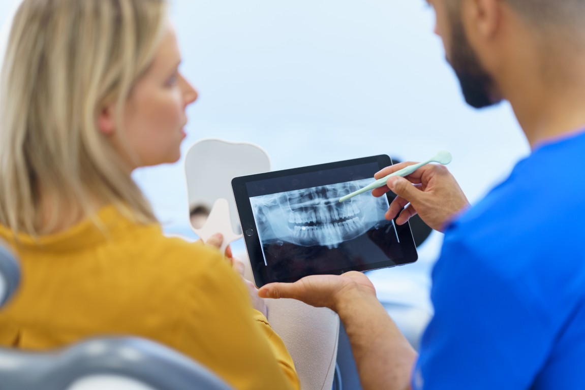
Digital Radiography is an advanced imaging technique that uses digital X-ray sensors instead of traditional photographic film to capture images of internal structures. This technology enables instant viewing of high-quality images on a computer screen, significantly reducing processing time and improving diagnostic accuracy. Digital Radiography offers numerous advantages, including reduced radiation exposure, easy storage and sharing of images, and the ability to enhance images for detailed evaluation. Widely utilized in medical, dental, veterinary, and industrial settings, Digital Radiography streamlines procedures, enhances efficiency, and ensures precise, reliable results for diagnostic and quality inspection purposes.
Panoramic Radiograph
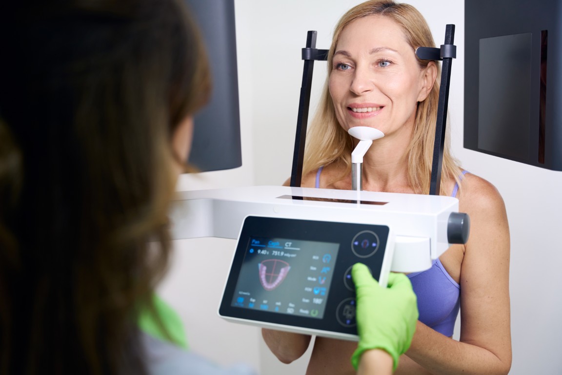
A Panoramic Radiograph, also known as a panoramic X-ray, is a specialized dental imaging technique that captures a wide, comprehensive view of the entire mouth, including the upper and lower jaws, teeth, temporomandibular joints (TMJ), sinuses, and surrounding structures. Unlike traditional dental X-rays that focus on specific teeth or small areas, Panoramic Radiography uses a rotating mechanism that moves around the patient's head to produce a single continuous image. This technology is essential in dentistry for evaluating tooth development, identifying impacted teeth, assessing jaw alignment, detecting cysts, tumors, or fractures, and planning treatments such as orthodontics, dental implants, and oral surgery. Panoramic Radiographs offer clinicians valuable diagnostic insights through a convenient, quick, and comfortable imaging procedure.
Cone Beam Computed Tomography
Cone Beam Computed Tomography (CBCT) is an advanced medical imaging technology that provides highly detailed three-dimensional views of anatomical structures, particularly in dental, oral surgery, and maxillofacial applications. Unlike traditional CT scans that use a fan-shaped beam, CBCT employs a cone-shaped X-ray beam that captures numerous images in a single rotation around the patient's head, significantly reducing radiation exposure and scan time. The collected images are digitally reconstructed into precise 3D models, enabling clinicians to accurately diagnose, plan treatments, and visualize complex structures such as teeth, bones, nerves, and soft tissues. CBCT has become an invaluable tool in dentistry and orthodontics, enhancing the safety, accuracy, and efficiency of various procedures, including implant planning, root canal treatment, orthodontic assessments, and oral surgeries.